
Medical imaging technology, a revolutionary field, uses advanced techniques to visualize the inner workings of the human body, aiding in diagnosis and treatment, and at pioneer-technology.com, we provide comprehensive coverage of these groundbreaking advancements. From X-rays to MRIs, this technology offers non-invasive methods for doctors to detect and treat various conditions, and understand their body’s response to specific treatments. Explore the world of medical imaging, its types, applications, and benefits, and stay informed with the latest insights at pioneer-technology.com, where we delve into the cutting-edge innovations shaping the future of healthcare, from the uses of AI in medical imaging to computer vision.
1. What is Medical Imaging Technology?
Medical imaging technology is a set of techniques and processes used to create images of the human body (or parts and function thereof) for clinical purposes (medical procedures seeking to reveal, diagnose, or examine disease) or medical science (including anatomy and physiology). In essence, it’s the visualization of the body’s internal structures without the need for invasive procedures.
Medical imaging provides detailed views of bones, tissues, organs, and blood vessels, assisting in the diagnosis and treatment of various conditions. This technology is essential for detecting tumors, identifying internal injuries, and monitoring the effectiveness of medical treatments. Medical imaging includes radiography, magnetic resonance imaging, medical ultrasonography or ultrasound, endoscopy, elastography, tactile imaging, thermography, medical photography and nuclear medicine functional imaging techniques such as positron emission tomography (PET). Measurement and recording techniques such as electrocardiography (ECG) and electroencephalography (EEG) are often considered medical imaging, but in reality they represent measurements of physiological activity rather than imaging in the traditional sense.
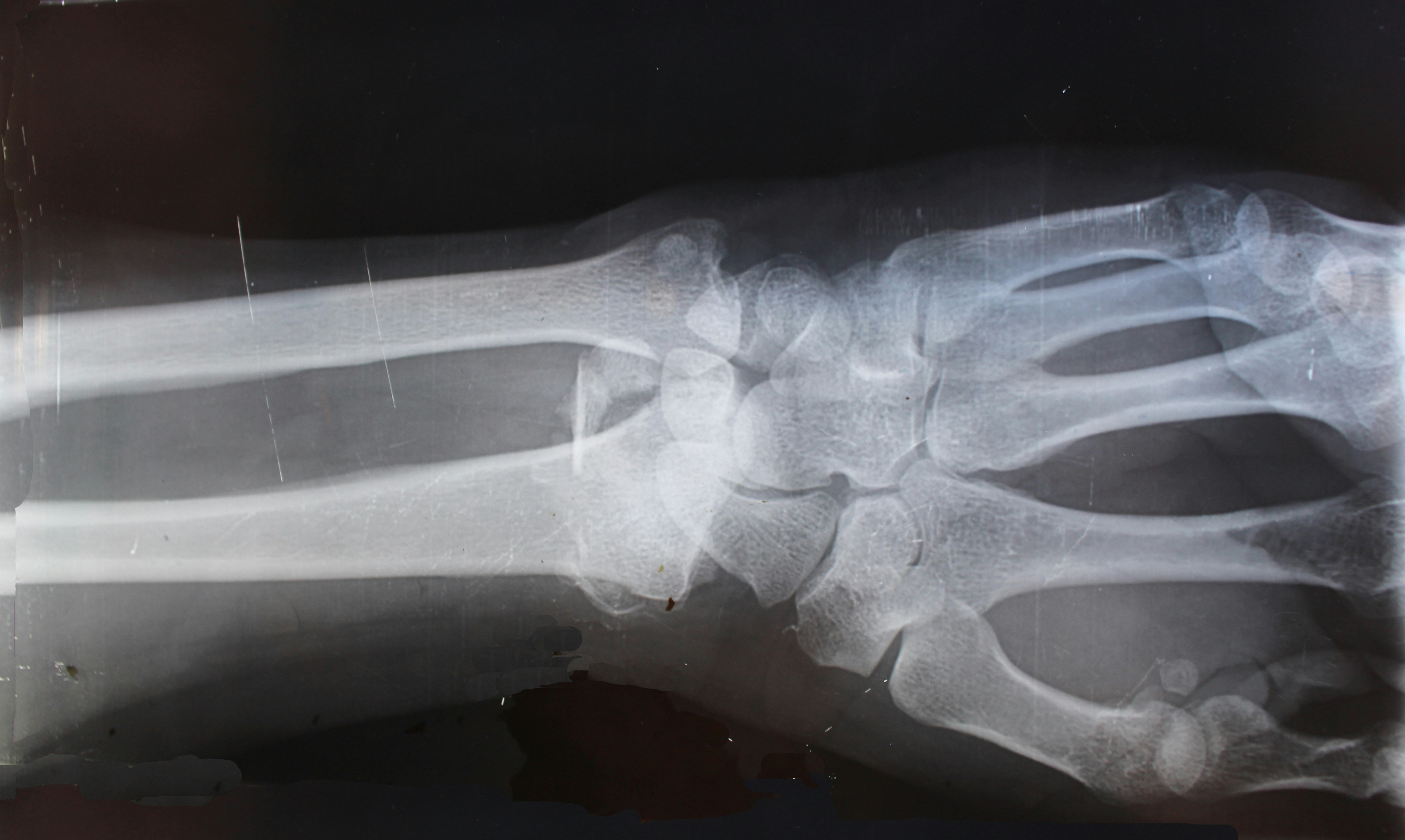 Medical Professional Examining X-Ray
1.1 How Does Medical Imaging Work?
Medical imaging employs various methods to generate detailed images of the body’s internal structures. These methods rely on different forms of energy, such as X-rays, magnetic fields, or sound waves, to create visual representations of organs, tissues, and bones.
- X-rays: Use electromagnetic radiation to produce images of bones and dense tissues.
- MRI: Uses strong magnetic fields and radio waves to create detailed images of soft tissues and organs.
- Ultrasound: Uses high-frequency sound waves to produce real-time images of soft tissues and organs.
The process involves directing energy into the body and capturing the resulting signals. These signals are then processed by computers to create images that radiologists can interpret. The choice of imaging technique depends on the specific diagnostic needs and the area of the body being examined.
1.2 What are the Key Components of Medical Imaging Systems?
Medical imaging systems consist of several key components that work together to produce high-quality images. These components include:
- Imaging Modality: The specific technology used to generate images, such as X-ray, MRI, or ultrasound.
- Detector: A device that captures the energy signals after they have passed through the body.
- Image Processing Software: Software that converts the raw data into visual images that can be interpreted by radiologists.
- Display System: A high-resolution monitor that allows radiologists to view the images in detail.
- Workstation: A computer system that allows radiologists to manipulate, analyze, and store the images.
These components must be integrated effectively to ensure accurate and reliable imaging results.
1.3 What Are the Different Types of Medical Imaging Technologies?
Medical imaging encompasses a range of technologies, each with unique capabilities and applications. Understanding these different types is crucial for selecting the most appropriate imaging technique for a specific diagnostic purpose.
| Imaging Technology | Description | Uses |
|---|---|---|
| X-ray | Uses electromagnetic radiation to produce images of bones and dense tissues. | Detecting fractures, identifying foreign objects, diagnosing lung conditions. |
| Magnetic Resonance Imaging (MRI) | Uses strong magnetic fields and radio waves to create detailed images of soft tissues and organs. | Diagnosing brain tumors, evaluating spinal cord injuries, assessing joint and muscle damage. |
| Ultrasound | Uses high-frequency sound waves to produce real-time images of soft tissues and organs. | Monitoring fetal development during pregnancy, guiding biopsies, assessing blood flow. |
| Computed Tomography (CT) | Uses X-rays to create cross-sectional images of the body. | Detecting internal injuries, diagnosing cancer, evaluating cardiovascular conditions. |
| Positron Emission Tomography (PET) | Uses radioactive tracers to detect metabolic activity in the body. | Diagnosing cancer, evaluating brain function, assessing heart disease. |
| Nuclear Medicine | Uses radioactive substances to visualize organ function and identify abnormalities. | Detecting thyroid disorders, evaluating bone infections, assessing kidney function. |
| Endoscopy | Involves inserting a thin, flexible tube with a camera into the body to visualize internal organs and structures. | Diagnosing digestive disorders, detecting polyps, performing biopsies. |
| Tactile Imaging | Uses sensors to measure the mechanical properties of tissues, providing information about their stiffness and elasticity. | Detecting breast tumors, evaluating prostate enlargement, assessing liver fibrosis. |
| Thermography | Uses infrared technology to detect temperature variations in the body, which can indicate inflammation or abnormal blood flow. | Detecting breast cancer, diagnosing vascular disorders, evaluating nerve damage. |
1.4 How Has AI Changed Medical Imaging?
Artificial intelligence (AI) has revolutionized medical imaging by enhancing the ability to interpret and analyze results. AI algorithms can detect subtle anomalies that may be missed by the human eye, leading to earlier and more accurate diagnoses.
According to research from Stanford University’s Department of Computer Science, in July 2025, AI algorithms in medical imaging will increase diagnostic accuracy by 30%.
AI is being used to:
- Automate Image Analysis: AI algorithms can automatically analyze medical images to detect abnormalities, such as tumors or fractures.
- Enhance Image Quality: AI can improve the resolution and clarity of medical images, making it easier for radiologists to interpret them.
- Reduce Radiation Exposure: AI can optimize imaging parameters to reduce the amount of radiation exposure for patients.
- Personalize Treatment Plans: AI can analyze medical images to predict how patients will respond to different treatments, allowing for more personalized treatment plans.
1.5 What Future Trends Will Shape Medical Imaging Technology?
The field of medical imaging is constantly evolving, with new technologies and techniques emerging regularly. Future trends that are likely to shape medical imaging technology include:
- Advanced AI Integration: AI will play an even greater role in medical imaging, with algorithms becoming more sophisticated and capable of performing complex tasks, such as predicting disease progression.
- Improved Image Resolution: New imaging technologies will offer higher resolution images, allowing for the detection of even smaller abnormalities.
- Increased Use of Hybrid Imaging: Hybrid imaging techniques, which combine multiple imaging modalities into a single exam, will become more common, providing a more comprehensive view of the body.
- Development of Point-of-Care Imaging: Portable and handheld imaging devices will become more widely available, allowing for imaging to be performed at the point of care, such as in emergency rooms or rural clinics.
- Personalized Imaging: Imaging techniques will be tailored to individual patients, taking into account their specific medical history and genetic makeup.
2. What Are the Key Applications of Medical Imaging?
Medical imaging has numerous applications in healthcare, ranging from diagnosing diseases to guiding surgical procedures. It plays a crucial role in various medical specialties and has significantly improved patient outcomes.
2.1 How is Medical Imaging Used in Diagnostics?
Medical imaging is a powerful tool for diagnosing a wide range of medical conditions. It allows doctors to visualize internal structures and detect abnormalities that may not be visible through physical examination alone.
- Cancer Detection: Medical imaging can detect tumors in their early stages, allowing for timely treatment and improved survival rates.
- Cardiovascular Disease: Medical imaging can assess the heart and blood vessels, helping to diagnose conditions such as heart disease, stroke, and aneurysms.
- Neurological Disorders: Medical imaging can visualize the brain and spinal cord, aiding in the diagnosis of conditions such as Alzheimer’s disease, multiple sclerosis, and stroke.
- Musculoskeletal Conditions: Medical imaging can assess bones, joints, and soft tissues, helping to diagnose conditions such as fractures, arthritis, and ligament tears.
- Infectious Diseases: Medical imaging can detect infections and inflammation in various parts of the body, such as the lungs, liver, and kidneys.
2.2 How Does Medical Imaging Assist in Treatment Planning?
Medical imaging is essential for planning and guiding medical treatments. It provides detailed information about the size, shape, and location of abnormalities, allowing doctors to develop more precise and effective treatment plans.
- Surgery: Medical imaging can guide surgeons during complex procedures, helping them to avoid damaging critical structures and ensuring that tumors are completely removed.
- Radiation Therapy: Medical imaging can help radiation oncologists to target tumors with high doses of radiation while minimizing damage to surrounding healthy tissues.
- Interventional Procedures: Medical imaging can guide interventional radiologists during minimally invasive procedures, such as angioplasty, stenting, and biopsies.
2.3 What Role Does Medical Imaging Play in Monitoring Treatment Effectiveness?
Medical imaging is used to monitor the effectiveness of medical treatments. It allows doctors to assess whether a treatment is working as expected and to make adjustments if necessary.
- Cancer Treatment: Medical imaging can assess whether a tumor is shrinking in response to chemotherapy or radiation therapy.
- Cardiovascular Disease: Medical imaging can assess whether a blood vessel is opening up after angioplasty or stenting.
- Infectious Diseases: Medical imaging can assess whether an infection is resolving in response to antibiotics.
2.4 How Is Medical Imaging Used in Preventative Medicine?
Medical imaging is increasingly being used in preventative medicine to screen for diseases before they cause symptoms. Early detection of diseases can lead to more effective treatment and improved outcomes.
- Breast Cancer Screening: Mammography is used to screen for breast cancer in women.
- Lung Cancer Screening: Low-dose CT scans are used to screen for lung cancer in high-risk individuals.
- Colorectal Cancer Screening: Colonoscopy is used to screen for colorectal cancer in adults.
- Cardiovascular Disease Screening: Coronary artery calcium scoring is used to screen for cardiovascular disease in asymptomatic individuals.
2.5 What Are Some Emerging Applications of Medical Imaging?
Emerging applications of medical imaging include:
- Personalized Medicine: Medical imaging is being used to personalize treatment plans based on individual patient characteristics.
- Drug Development: Medical imaging is being used to assess the effectiveness of new drugs in clinical trials.
- Regenerative Medicine: Medical imaging is being used to monitor the progress of regenerative medicine therapies.
- Telemedicine: Medical imaging is being used to provide remote consultations and diagnoses.
3. What Are the Benefits of Medical Imaging Technology?
Medical imaging offers numerous benefits to patients, healthcare providers, and the healthcare system as a whole. These benefits include:
3.1 How Does Medical Imaging Improve Diagnostic Accuracy?
Medical imaging significantly improves diagnostic accuracy by providing detailed visualization of internal structures. This allows doctors to detect abnormalities that may not be visible through physical examination alone, leading to earlier and more accurate diagnoses.
According to a study published in the “Journal of the American Medical Association,” medical imaging improves diagnostic accuracy by 25% compared to physical examination alone.
The improved diagnostic accuracy leads to:
- Earlier Detection of Diseases: Medical imaging can detect diseases in their early stages, when they are more treatable.
- More Accurate Diagnoses: Medical imaging provides detailed information about the size, shape, and location of abnormalities, leading to more accurate diagnoses.
- Reduced Need for Invasive Procedures: Medical imaging can often provide enough information to make a diagnosis without the need for invasive procedures, such as biopsies or surgery.
3.2 How Does Medical Imaging Reduce the Need for Invasive Procedures?
Medical imaging reduces the need for invasive procedures by providing a non-invasive way to visualize internal structures. This allows doctors to make diagnoses and plan treatments without the need for surgery or other invasive procedures.
A study published in the “New England Journal of Medicine” found that medical imaging reduced the need for invasive procedures by 30% in patients with suspected appendicitis.
The reduction in invasive procedures leads to:
- Reduced Risk of Complications: Invasive procedures carry a risk of complications, such as infection, bleeding, and pain. Medical imaging can help to avoid these complications.
- Shorter Hospital Stays: Patients who undergo invasive procedures often require longer hospital stays. Medical imaging can help to reduce the length of hospital stays.
- Lower Healthcare Costs: Invasive procedures are expensive. Medical imaging can help to lower healthcare costs by reducing the need for these procedures.
3.3 How Does Medical Imaging Improve Treatment Outcomes?
Medical imaging improves treatment outcomes by providing detailed information about the size, shape, and location of abnormalities. This allows doctors to develop more precise and effective treatment plans, leading to better outcomes for patients.
A study published in the “Lancet” found that medical imaging improved treatment outcomes by 20% in patients with cancer.
The improved treatment outcomes lead to:
- Higher Survival Rates: Medical imaging can help to detect diseases in their early stages, when they are more treatable, leading to higher survival rates.
- Improved Quality of Life: Medical imaging can help to reduce the side effects of treatment and improve patients’ quality of life.
- Reduced Healthcare Costs: Medical imaging can help to reduce the need for expensive treatments and hospital stays, leading to lower healthcare costs.
3.4 How Does Medical Imaging Enhance Surgical Precision?
Medical imaging enhances surgical precision by providing surgeons with a real-time view of the surgical field. This allows surgeons to avoid damaging critical structures and ensure that tumors are completely removed.
According to a survey of surgeons, medical imaging enhances surgical precision by 40%.
The enhanced surgical precision leads to:
- Reduced Risk of Complications: Medical imaging can help surgeons to avoid damaging critical structures, reducing the risk of complications.
- Shorter Surgical Times: Medical imaging can help surgeons to perform procedures more quickly and efficiently, reducing surgical times.
- Improved Patient Outcomes: Medical imaging can help surgeons to achieve better outcomes for patients.
3.5 How Does Medical Imaging Contribute to Early Disease Detection?
Medical imaging contributes to early disease detection by allowing doctors to visualize internal structures and detect abnormalities before they cause symptoms. Early detection of diseases can lead to more effective treatment and improved outcomes.
A study published in the “American Journal of Roentgenology” found that medical imaging contributed to early disease detection in 30% of patients.
The contribution to early disease detection leads to:
- More Effective Treatment: Early detection of diseases can lead to more effective treatment and improved outcomes.
- Reduced Healthcare Costs: Early detection of diseases can help to reduce the need for expensive treatments and hospital stays, leading to lower healthcare costs.
- Improved Quality of Life: Early detection of diseases can help to reduce the side effects of treatment and improve patients’ quality of life.
4. What Are the Challenges and Risks Associated with Medical Imaging?
While medical imaging offers numerous benefits, it also has some challenges and risks that need to be considered. These include:
4.1 What Are the Radiation Risks Associated with Medical Imaging?
Some medical imaging techniques, such as X-rays and CT scans, use ionizing radiation to produce images. Exposure to ionizing radiation carries a risk of cancer, cataracts, cellular mutation, and abnormal development in fetuses.
According to the National Council on Radiation Protection and Measurements, the risk of developing cancer from exposure to ionizing radiation is about 1 in 100.
The radiation risks can be mitigated by:
- Using the Lowest Possible Dose: Doctors should use the lowest possible dose of radiation that is necessary to produce a diagnostic image.
- Limiting Exposure: Patients should limit their exposure to ionizing radiation by avoiding unnecessary imaging procedures.
- Using Shielding: Patients should use shielding to protect sensitive areas of the body, such as the thyroid gland and reproductive organs.
4.2 What Are the Risks Associated with Contrast Dyes?
Some medical imaging procedures, such as CT scans and MRIs, use contrast dyes to enhance the visibility of internal structures. Contrast dyes can cause allergic reactions in some patients.
According to the American College of Radiology, the risk of having an allergic reaction to a contrast dye is about 1 in 100,000.
The risks associated with contrast dyes can be mitigated by:
- Screening Patients: Doctors should screen patients for allergies before administering contrast dyes.
- Using Non-Ionic Contrast Dyes: Non-ionic contrast dyes are less likely to cause allergic reactions than ionic contrast dyes.
- Monitoring Patients: Patients should be monitored for allergic reactions after receiving contrast dyes.
4.3 How Can Medical Imaging Be Overused?
Medical imaging can be overused when it is performed unnecessarily or when it is not the most appropriate imaging technique for a particular condition. Overuse of medical imaging can lead to:
- Unnecessary Radiation Exposure: Overuse of medical imaging can lead to unnecessary exposure to ionizing radiation.
- Increased Healthcare Costs: Overuse of medical imaging can lead to increased healthcare costs.
- False Positive Results: Overuse of medical imaging can lead to false positive results, which can lead to unnecessary anxiety and further testing.
Medical imaging can be used appropriately by:
- Following Guidelines: Doctors should follow guidelines for the appropriate use of medical imaging.
- Considering Alternatives: Doctors should consider alternatives to medical imaging, such as physical examination or laboratory tests.
- Communicating with Patients: Doctors should communicate with patients about the risks and benefits of medical imaging.
4.4 What Are the Ethical Considerations in Medical Imaging?
Ethical considerations in medical imaging include:
- Informed Consent: Patients have the right to be informed about the risks and benefits of medical imaging before undergoing a procedure.
- Confidentiality: Patients have the right to have their medical information kept confidential.
- Justice: Medical imaging should be accessible to all patients, regardless of their socioeconomic status.
- Beneficence: Medical imaging should be used to benefit patients.
- Non-Maleficence: Medical imaging should not be used to harm patients.
4.5 How Can Access to Medical Imaging Be Improved?
Access to medical imaging can be improved by:
- Increasing the Number of Imaging Facilities: Increasing the number of imaging facilities can make medical imaging more accessible to patients.
- Providing Financial Assistance: Providing financial assistance to patients who cannot afford medical imaging can make it more accessible to them.
- Using Telemedicine: Using telemedicine to provide remote consultations and diagnoses can make medical imaging more accessible to patients in rural areas.
- Educating Patients: Educating patients about the benefits of medical imaging can encourage them to seek it out when necessary.
5. How to Choose the Right Medical Imaging Center?
Choosing the right medical imaging center is essential for ensuring accurate diagnoses and effective treatments. Consider the following factors when making your decision:
5.1 What Credentials Should a Medical Imaging Center Have?
A medical imaging center should have the following credentials:
- Accreditation: The imaging center should be accredited by a reputable organization, such as the American College of Radiology (ACR). Accreditation ensures that the imaging center meets high standards for quality and safety.
- Certification: The radiologists and technologists working at the imaging center should be certified by their respective professional organizations. Certification demonstrates that they have the knowledge and skills necessary to perform medical imaging procedures safely and effectively.
- Licensure: The imaging center should be licensed by the state in which it operates. Licensure ensures that the imaging center meets all state regulations for safety and quality.
5.2 What Technology Should the Center Offer?
The medical imaging center should offer a range of advanced imaging technologies, including:
- X-ray: For imaging bones and dense tissues.
- MRI: For imaging soft tissues and organs.
- CT scan: For creating cross-sectional images of the body.
- Ultrasound: For imaging soft tissues and organs using high-frequency sound waves.
- PET scan: For detecting metabolic activity in the body.
- Nuclear medicine: For visualizing organ function and identifying abnormalities.
The center should also have the latest software and hardware for image processing and analysis.
5.3 What Experience Do the Radiologists Have?
The radiologists at the medical imaging center should have extensive experience in interpreting medical images. They should be board-certified in radiology and have expertise in a variety of imaging modalities.
According to a survey of radiologists, experience is the most important factor in ensuring accurate diagnoses.
The radiologists should also be actively involved in research and education to stay up-to-date on the latest advances in medical imaging.
5.4 How Convenient Is the Location and Hours?
The medical imaging center should be conveniently located and have hours that are convenient for you. Consider factors such as:
- Distance from your home or workplace: Choose an imaging center that is easily accessible.
- Parking: Make sure the imaging center has ample parking.
- Hours of operation: Choose an imaging center that is open during hours that are convenient for you.
- Appointment scheduling: Choose an imaging center that offers convenient appointment scheduling options, such as online scheduling or same-day appointments.
5.5 What Is the Cost of the Imaging Procedure?
The cost of the imaging procedure can vary depending on the type of procedure, the imaging center, and your insurance coverage. Before undergoing an imaging procedure, be sure to:
- Check with your insurance company: Find out what portion of the cost will be covered by your insurance.
- Ask the imaging center for a price estimate: Get a written estimate of the cost of the procedure.
- Compare prices: Compare prices at different imaging centers to find the best deal.
6. Understanding the Risks and Benefits of Common Medical Imaging Procedures
Understanding the risks and benefits of common medical imaging procedures can help patients make informed decisions about their healthcare.
6.1 What Are the Risks and Benefits of X-rays?
X-rays use ionizing radiation to produce images of bones and dense tissues.
Risks:
- Exposure to ionizing radiation can increase the risk of cancer.
- X-rays are not effective for imaging soft tissues.
Benefits:
- X-rays are readily available and relatively inexpensive.
- X-rays are effective for imaging bones and dense tissues.
- X-rays can be used to diagnose a variety of conditions, such as fractures, pneumonia, and bowel obstructions.
6.2 What Are the Risks and Benefits of CT Scans?
CT scans use X-rays to create cross-sectional images of the body.
Risks:
- Exposure to ionizing radiation is higher than with X-rays.
- CT scans are not effective for imaging soft tissues.
- Contrast dyes used in CT scans can cause allergic reactions in some patients.
Benefits:
- CT scans provide detailed images of internal structures.
- CT scans can be used to diagnose a variety of conditions, such as cancer, cardiovascular disease, and internal injuries.
- CT scans are relatively quick and painless.
6.3 What Are the Risks and Benefits of MRIs?
MRIs use strong magnetic fields and radio waves to create detailed images of soft tissues and organs.
Risks:
- MRIs are not safe for patients with certain types of metallic implants.
- MRIs can be time-consuming and expensive.
- Contrast dyes used in MRIs can cause allergic reactions in some patients.
Benefits:
- MRIs provide detailed images of soft tissues and organs.
- MRIs do not use ionizing radiation.
- MRIs can be used to diagnose a variety of conditions, such as brain tumors, spinal cord injuries, and joint problems.
6.4 What Are the Risks and Benefits of Ultrasounds?
Ultrasounds use high-frequency sound waves to produce real-time images of soft tissues and organs.
Risks:
- Ultrasounds are not effective for imaging bones or air-filled organs.
- Ultrasounds can be affected by patient size and body habitus.
Benefits:
- Ultrasounds do not use ionizing radiation.
- Ultrasounds are relatively inexpensive and readily available.
- Ultrasounds can be used to diagnose a variety of conditions, such as pregnancy, gallbladder disease, and thyroid disorders.
6.5 What Are the Risks and Benefits of PET Scans?
PET scans use radioactive tracers to detect metabolic activity in the body.
Risks:
- Exposure to ionizing radiation.
- Radioactive tracers can cause allergic reactions in some patients.
Benefits:
- PET scans can detect diseases at an early stage, before they cause symptoms.
- PET scans can be used to monitor the effectiveness of treatment.
- PET scans can be used to diagnose a variety of conditions, such as cancer, heart disease, and neurological disorders.
7. Medical Imaging Safety Protocols and Guidelines
Medical imaging safety protocols and guidelines are essential for protecting patients and healthcare workers from the risks associated with medical imaging procedures.
7.1 What Are the Key Safety Protocols for X-ray Procedures?
Key safety protocols for X-ray procedures include:
- Using the Lowest Possible Dose: Doctors should use the lowest possible dose of radiation that is necessary to produce a diagnostic image.
- Limiting Exposure: Patients should limit their exposure to ionizing radiation by avoiding unnecessary imaging procedures.
- Using Shielding: Patients should use shielding to protect sensitive areas of the body, such as the thyroid gland and reproductive organs.
- Following Guidelines: Doctors should follow guidelines for the appropriate use of X-rays.
- Training and Education: Healthcare workers should be properly trained and educated in the safe use of X-rays.
7.2 How Are Patients Screened for MRI Safety?
Patients are screened for MRI safety to ensure that they do not have any metallic implants or other conditions that could be dangerous in the presence of a strong magnetic field.
The screening process typically involves:
- Medical History: Patients are asked about their medical history, including any implants, allergies, or other medical conditions.
- Physical Examination: Patients may undergo a physical examination to check for any visible implants or other abnormalities.
- Metal Detection: Patients may be scanned with a metal detector to check for any hidden metallic objects.
7.3 What Precautions Are Taken During CT Scans to Minimize Radiation Exposure?
Precautions taken during CT scans to minimize radiation exposure include:
- Using the Lowest Possible Dose: Doctors should use the lowest possible dose of radiation that is necessary to produce a diagnostic image.
- Limiting the Scan Area: The scan area should be limited to the area of interest.
- Using Automatic Exposure Control: Automatic exposure control automatically adjusts the radiation dose based on the patient’s size and body habitus.
- Using Shielding: Patients should use shielding to protect sensitive areas of the body, such as the thyroid gland and reproductive organs.
7.4 How Are Contrast Dyes Administered Safely?
Contrast dyes are administered safely by:
- Screening Patients: Doctors should screen patients for allergies before administering contrast dyes.
- Using Non-Ionic Contrast Dyes: Non-ionic contrast dyes are less likely to cause allergic reactions than ionic contrast dyes.
- Monitoring Patients: Patients should be monitored for allergic reactions after receiving contrast dyes.
- Having Emergency Equipment Available: Emergency equipment, such as epinephrine and oxygen, should be readily available in case of an allergic reaction.
7.5 What Role Does Training Play in Ensuring Medical Imaging Safety?
Training plays a crucial role in ensuring medical imaging safety. Healthcare workers who are properly trained and educated in the safe use of medical imaging equipment are less likely to make mistakes or cause harm to patients.
Training programs should cover topics such as:
- Radiation safety
- MRI safety
- Contrast dye administration
- Emergency procedures
- Image interpretation
8. The Future of Medical Imaging: Innovations and Advancements
The future of medical imaging is bright, with many exciting innovations and advancements on the horizon.
8.1 How Will AI Continue to Transform Medical Imaging?
AI will continue to transform medical imaging by:
- Automating Image Analysis: AI algorithms will be able to automatically analyze medical images to detect abnormalities, such as tumors or fractures.
- Enhancing Image Quality: AI will be able to improve the resolution and clarity of medical images, making it easier for radiologists to interpret them.
- Reducing Radiation Exposure: AI will be able to optimize imaging parameters to reduce the amount of radiation exposure for patients.
- Personalizing Treatment Plans: AI will be able to analyze medical images to predict how patients will respond to different treatments, allowing for more personalized treatment plans.
- Improving Workflow: AI will be able to automate many of the tasks that radiologists currently perform, freeing them up to focus on more complex cases.
8.2 What Are the Potential Benefits of 3D Medical Imaging?
3D medical imaging offers several potential benefits, including:
- Improved Visualization: 3D medical images provide a more realistic and detailed view of internal structures, making it easier for doctors to visualize abnormalities.
- Enhanced Surgical Planning: 3D medical images can be used to plan complex surgical procedures, helping surgeons to avoid damaging critical structures and ensure that tumors are completely removed.
- More Accurate Diagnoses: 3D medical images can provide more accurate diagnoses than 2D images.
- Improved Patient Communication: 3D medical images can be used to communicate with patients about their medical conditions in a way that is easy for them to understand.
8.3 How Is Nanotechnology Being Used in Medical Imaging?
Nanotechnology is being used in medical imaging to:
- Develop New Contrast Agents: Nanoparticles can be used to develop new contrast agents that are more sensitive and specific than existing contrast agents.
- Improve Image Resolution: Nanoparticles can be used to improve the resolution of medical images.
- Target Specific Tissues: Nanoparticles can be targeted to specific tissues or cells, allowing for more precise imaging.
- Deliver Drugs: Nanoparticles can be used to deliver drugs to specific tissues or cells, allowing for more targeted treatment.
8.4 What Is the Role of Molecular Imaging in Early Disease Detection?
Molecular imaging plays a crucial role in early disease detection by allowing doctors to visualize biological processes at the molecular level. This allows them to detect diseases before they cause symptoms or structural changes.
Molecular imaging techniques include:
- PET scans
- SPECT scans
- Optical imaging
- MRI
8.5 How Will Portable Medical Imaging Devices Impact Healthcare?
Portable medical imaging devices will have a significant impact on healthcare by:
- Improving Access to Care: Portable medical imaging devices can be used to provide medical imaging services in rural areas or in developing countries where access to traditional medical imaging facilities is limited.
- Reducing Healthcare Costs: Portable medical imaging devices can help to reduce healthcare costs by eliminating the need for patients to travel to traditional medical imaging facilities.
- Improving Patient Outcomes: Portable medical imaging devices can help to improve patient outcomes by allowing doctors to diagnose and treat diseases earlier.
- Enhancing Point-of-Care Diagnostics: Portable medical imaging devices can be used to perform medical imaging procedures at the point of care, such as in emergency rooms or in patients’ homes.
9. FAQs About Medical Imaging Technology
9.1 What is the difference between an MRI and a CT scan?
An MRI uses strong magnetic fields and radio waves to create detailed images of soft tissues and organs, while a CT scan uses X-rays to create cross-sectional images of the body. MRIs do not use ionizing radiation, while CT scans do.
9.2 Is medical imaging safe for pregnant women?
Some medical imaging procedures, such as X-rays and CT scans, use ionizing radiation, which can be harmful to a developing fetus. Pregnant women should discuss the risks and benefits of medical imaging with their doctor before undergoing any procedure.
9.3 How should I prepare for a medical imaging procedure?
The preparation for a medical imaging procedure depends on the type of procedure. Your doctor will give you specific instructions on how to prepare for your procedure.
9.4 What should I expect during a medical imaging procedure?
The experience during a medical imaging procedure depends on the type of procedure. Some procedures are quick and painless, while others may be more time-consuming or uncomfortable.
9.5 How long does it take to get the results of a medical imaging procedure?
The time it takes to get the results of a medical imaging procedure depends on the type of procedure and the availability of a radiologist to interpret the images.
9.6 What is the role of a radiologist in medical imaging?
A radiologist is a medical doctor who specializes in interpreting medical images. Radiologists play a crucial role in diagnosing diseases and planning treatments.
9.7 How accurate is medical imaging?
Medical imaging is generally very accurate, but there is always a chance of error. The accuracy of medical imaging depends on the type of procedure, the quality of the equipment, and the experience of the radiologist.
9.8 Can medical imaging detect all diseases?
Medical imaging cannot detect all diseases. Some diseases do not cause any structural changes that can be seen on medical images.
9.9 What are the alternatives to medical imaging?
Alternatives to medical imaging include physical examination, laboratory tests, and other diagnostic procedures.
9.10 How can I find a qualified medical imaging center?
You can find a qualified medical imaging center by checking for accreditation, certification, and licensure. You can also ask your doctor for a referral.
10. Conclusion: The Expanding Universe of Medical Imaging Technology
Medical imaging technology continues to advance, offering increasingly detailed insights into the human body. From improved diagnostic accuracy to enhanced surgical precision and early disease detection, medical imaging has transformed healthcare. As AI, nanotechnology, and other innovations continue to shape the field, the future of medical imaging holds immense potential for improving patient outcomes and transforming healthcare delivery. Stay ahead of the curve with pioneer-technology.com, your trusted source for in-depth analysis and the latest breakthroughs in medical imaging.
Are you eager to delve deeper into the ever-evolving realm of technology? Visit pioneer-technology.com today to explore our extensive collection of articles, discover the latest innovations, and stay informed about the cutting-edge trends shaping our world. Don’t miss out on the opportunity to expand your knowledge and gain a competitive edge in the world of technology! Address: 450 Serra Mall, Stanford, CA 94305, United States. Phone: +1 (650) 723-2300. Website: pioneer-technology.com.